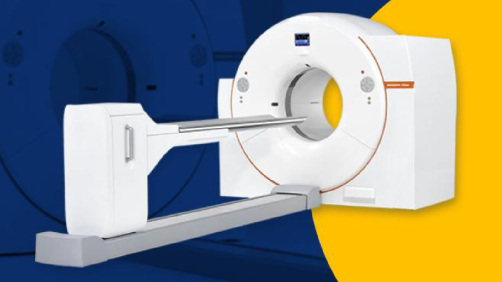
A positron emission tomography scan is a diagnostic imaging technique in the field of nuclear medicine. The PET scan operates with a radioactive dye or contrast known as FDG (fluorodeoxyglucose). The contrast agent has glucose or sugar in it which is utilised by the cells of the body. In abnormally functioning cells of the body, metabolic activity is raised. The contrast is absorbed more by the more active cells, thereby presenting them more clearly in the scan images of the scan. The CT scan (Computed tomography) is a diagnostic technique that operates on the ionising radiations. PET and CT scans are used as a hybrid imaging technique that combines the advantages of both the PET and CT scan, where PET scan if efficient in determining even the slightest metabolic changes in the body cells or the tissues, and the CT scan produces cross-sectional detailed and high contrast images of the body in form of slices. When the detailed images are combined and collaborated with their metabolic functioning, a hybrid PET-CT scan is born.
The contrast agent FDG when used with the PET-CT scan can be used to
- Pinpoint the location of the cancer cells
- Plan the suitable treatment plan
- To measure the effectiveness of the ongoing treatment
- Evaluate and identify the recurrence of cancer
The CT scan is conducted along with the PET study as a hybrid PET-CT technique.
The radioactive tracer used in the PET-CT scanning can be administered in your body through various methods. Once the scanner is administered and absorbed by your body cells, the tracer gets absorbed by the body tissue cells that absorb the contrast. Once the contrast is absorbed, the scanner can identify the abnormally functioning cells.
The contrast can be administered by various routes including the most commonly used IV route, and the oral route. In the IV or intravenous route, the contrast is administered into your arm, hand, or through the central venous catheter. You can also be suggested to swallow the contrast to better appreciate certain GI (gastrointestinal) organs. When the contrast is administered through an oral route, the contrast is prepared as a solution. The oral contrast agent can be iodinated contrast that contains iodine as a component. Or it can be diluted barium-based contrast sweetened with saccharin.
The contrast needs to be administered at least 30-60 minutes before the scan is done,
The contrast where iodine-based and non-iodine-based, can have certain side effects. Most likely an allergic reaction is the side effect that a contrast agent contains in themselves. Iodine based contrast dye has more potential to pose an allergic reaction, while a non iodine based contrast agent like gadolinium contrast dye has less potential to cause allergic reaction. The best diagnostic centre uses the contrast agent that has least potential to cause allergic reactions.
Add a Comment